
MARKETICA_PREVIEW/00-marketica-preview-sale37.jpg
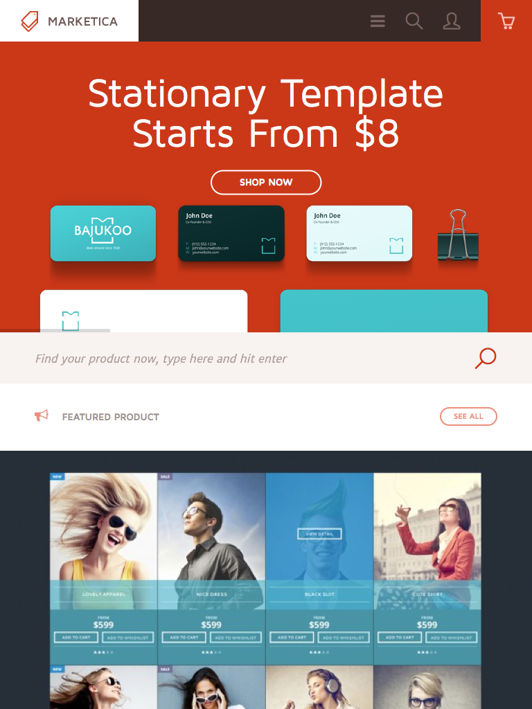
MARKETICA_PREVIEW/01_marketica2_homepage.png
MARKETICA_PREVIEW/02_marketica2_shop_page.png
MARKETICA_PREVIEW/03_marketica2_single_product_page.png
MARKETICA_PREVIEW/04_marketica2_cart_page.png
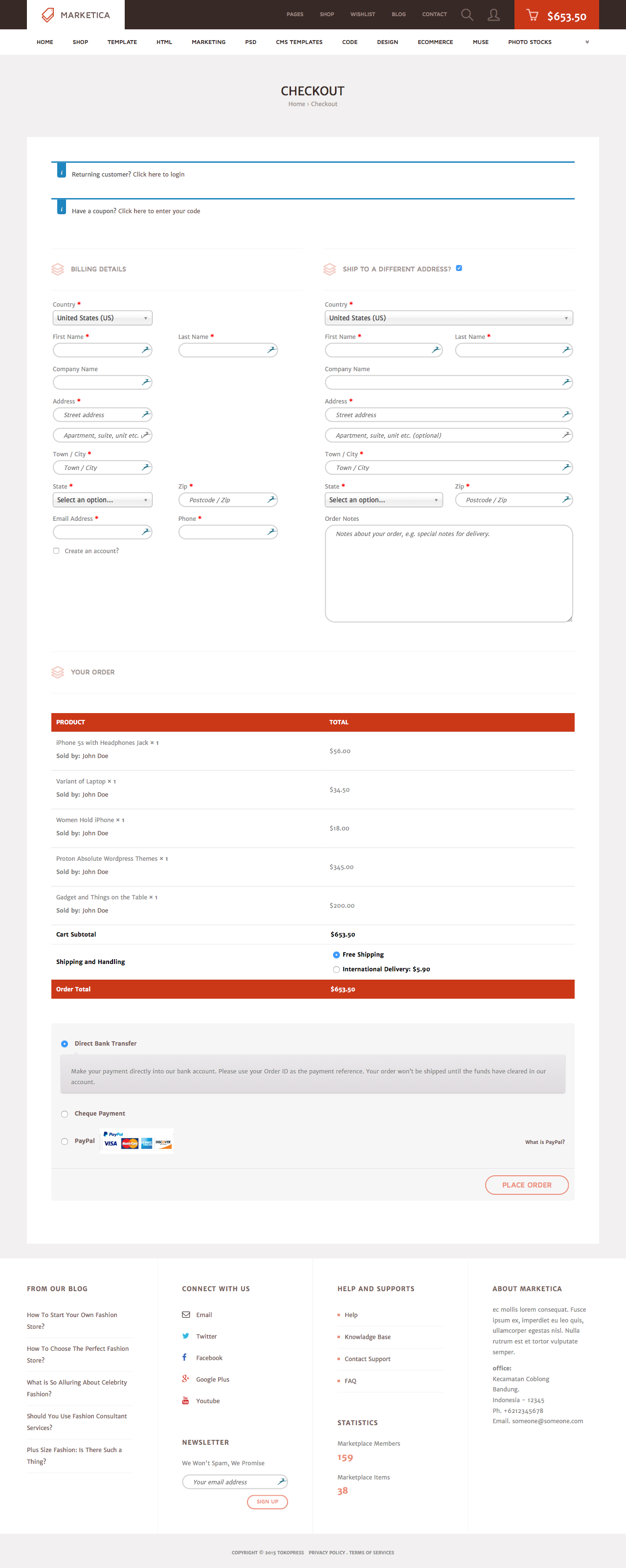
MARKETICA_PREVIEW/05_marketica2_checkout_page.png
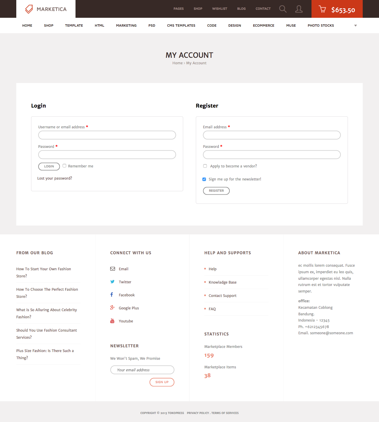
MARKETICA_PREVIEW/06_marketica2_myaccount_login_page.png
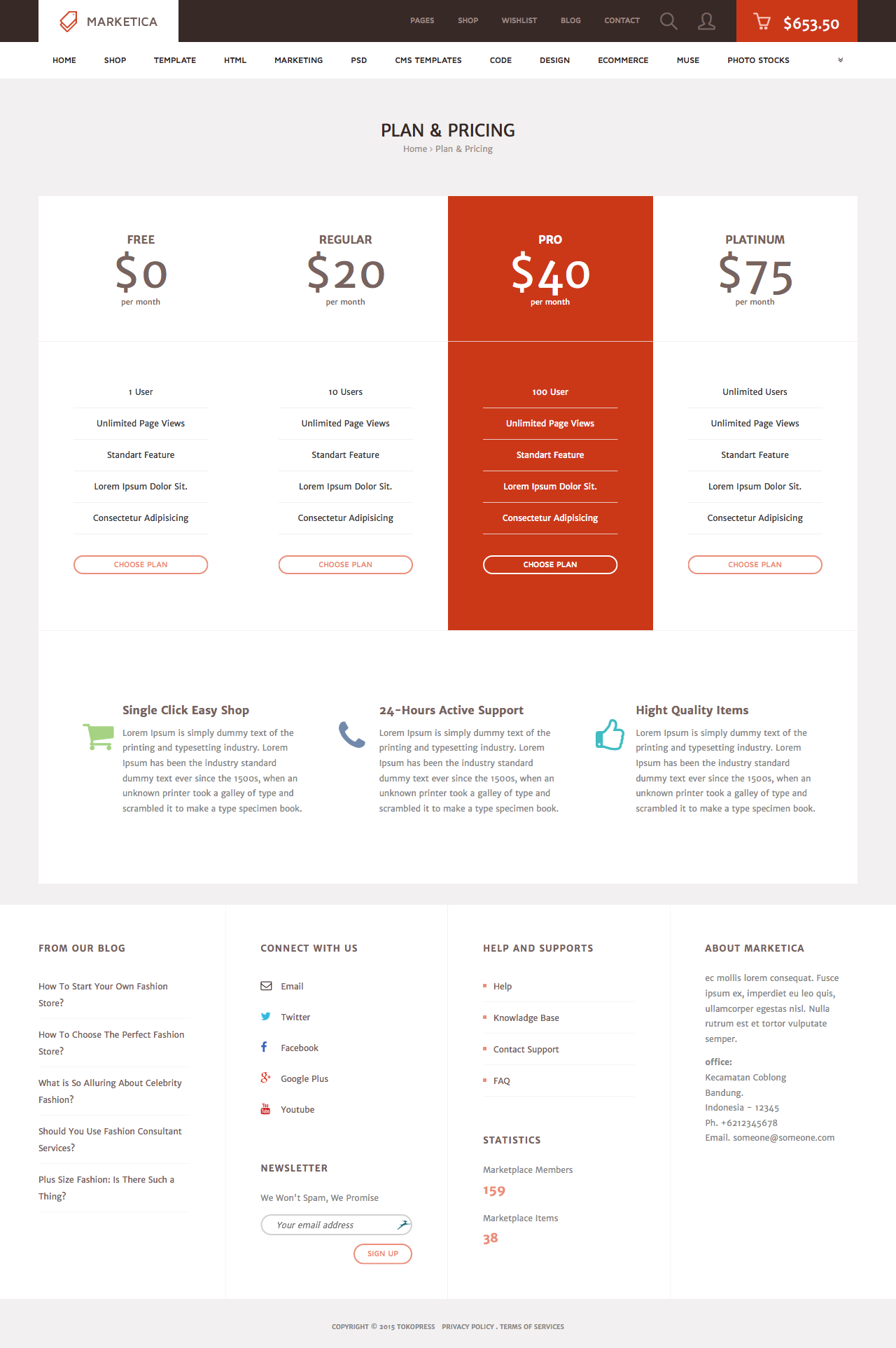
MARKETICA_PREVIEW/07_marketica2_plan_and_pricing_page.png
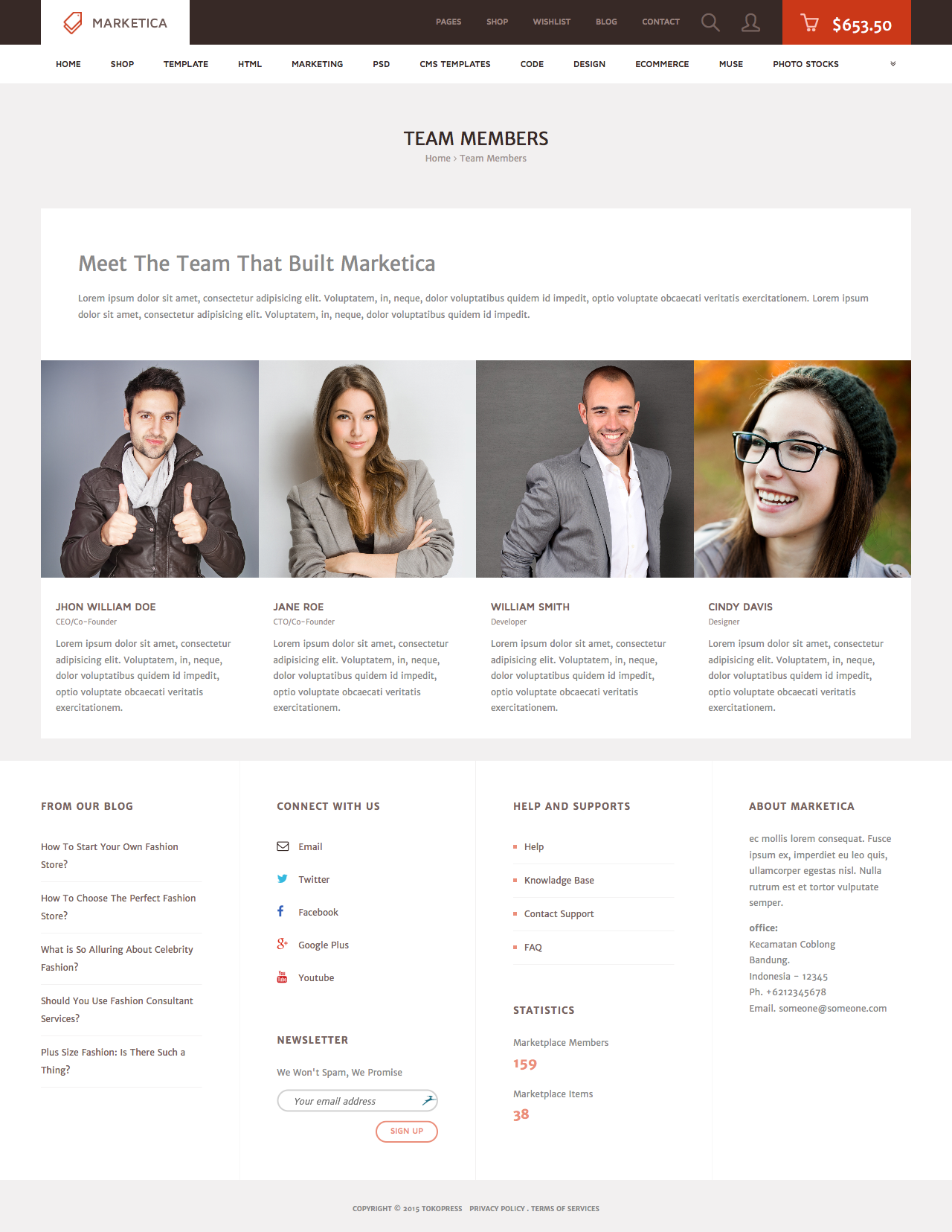
MARKETICA_PREVIEW/08_marketica2_team_members_page.png
MARKETICA_PREVIEW/09_marketica2_contact_page_template.png
MARKETICA_PREVIEW/10_marketica2_blog_page.png
MARKETICA_PREVIEW/11_marketica2_blog_post_formats.png
MARKETICA_PREVIEW/12_marketica2_single_product_page.png
MARKETICA_PREVIEW/13_marketica2_theme_customizer.png
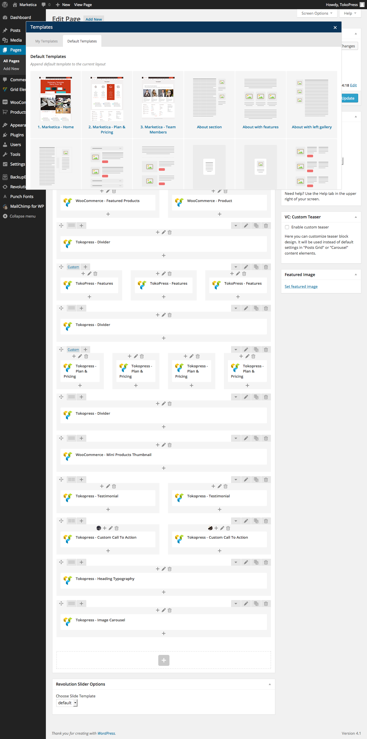
MARKETICA_PREVIEW/14_marketica2_visualcomposer_templates.png
MARKETICA_PREVIEW/15_marketica2_tablet_view.png
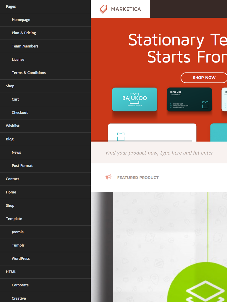
MARKETICA_PREVIEW/16_marketica2_tablet_view_offcanvas_menu.png
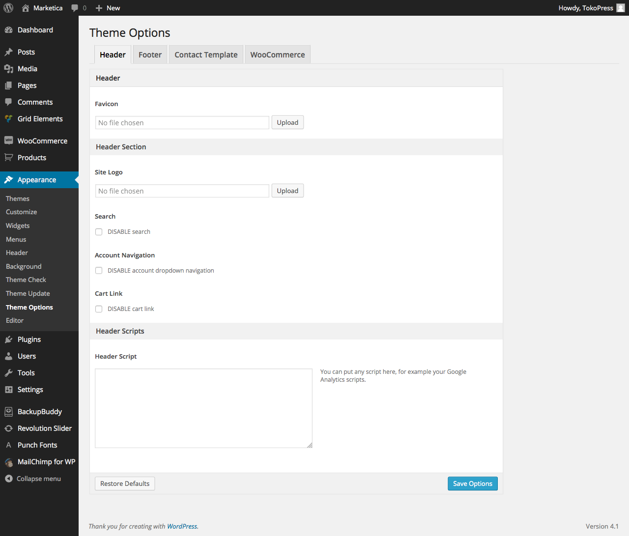
MARKETICA_PREVIEW/17_marketica2_themeoptions_header.png
MARKETICA_PREVIEW/18_marketica2_themeoptions_footer.png
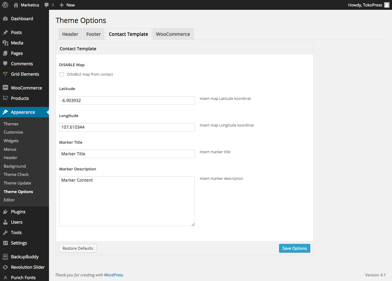
MARKETICA_PREVIEW/19_marketica2_themeoptions_contact.png
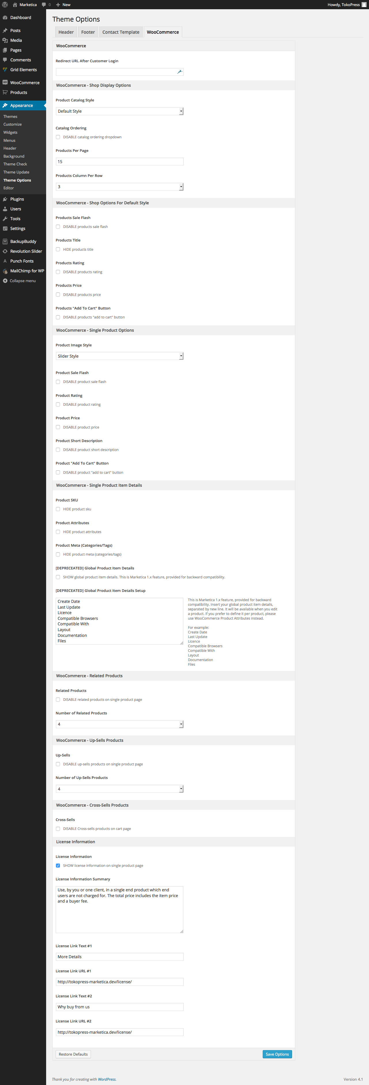
MARKETICA_PREVIEW/20_marketica2_themeoptions_woocommerce.png
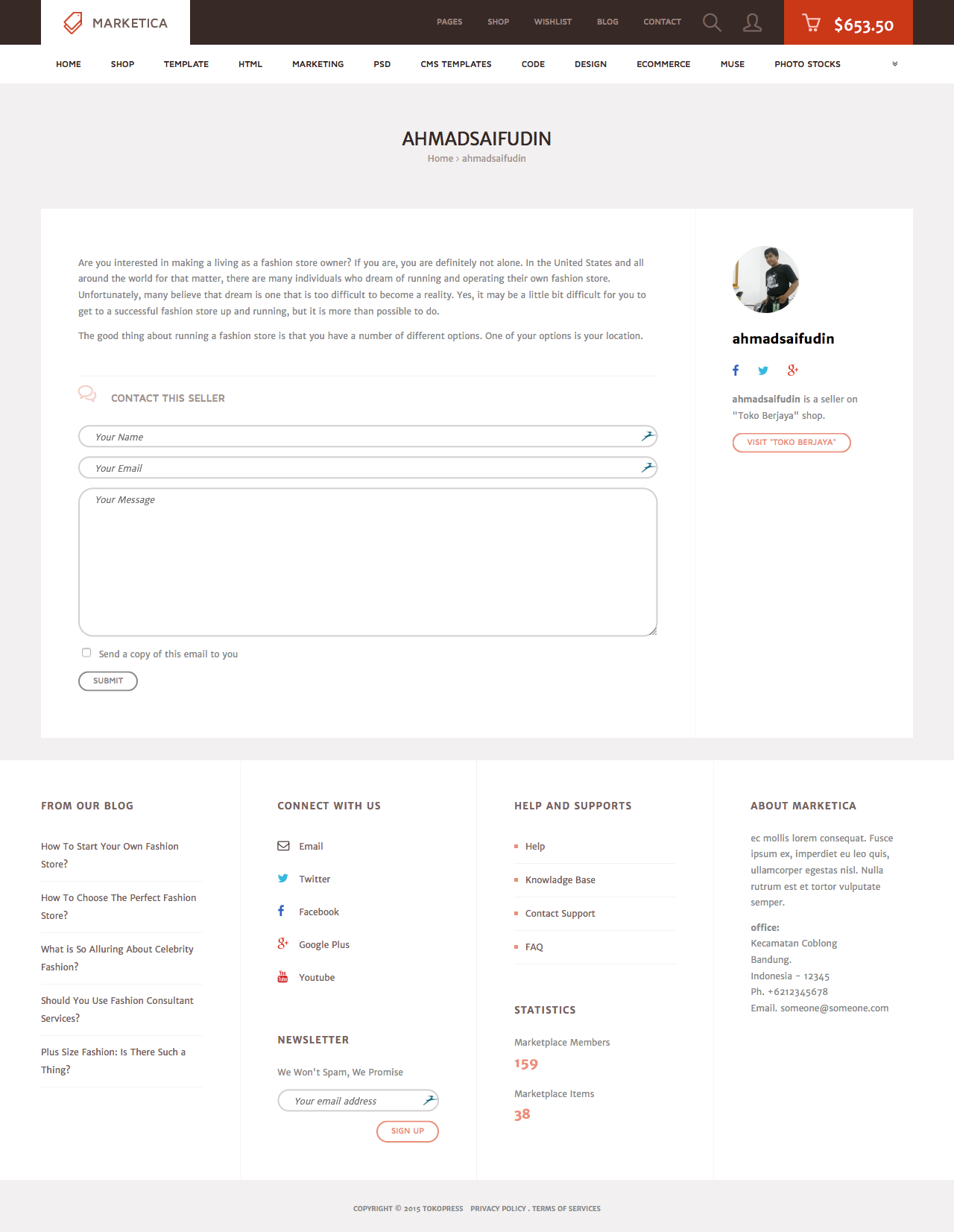
MARKETICA_PREVIEW/21_marketica2_wcvendors_user_page.png
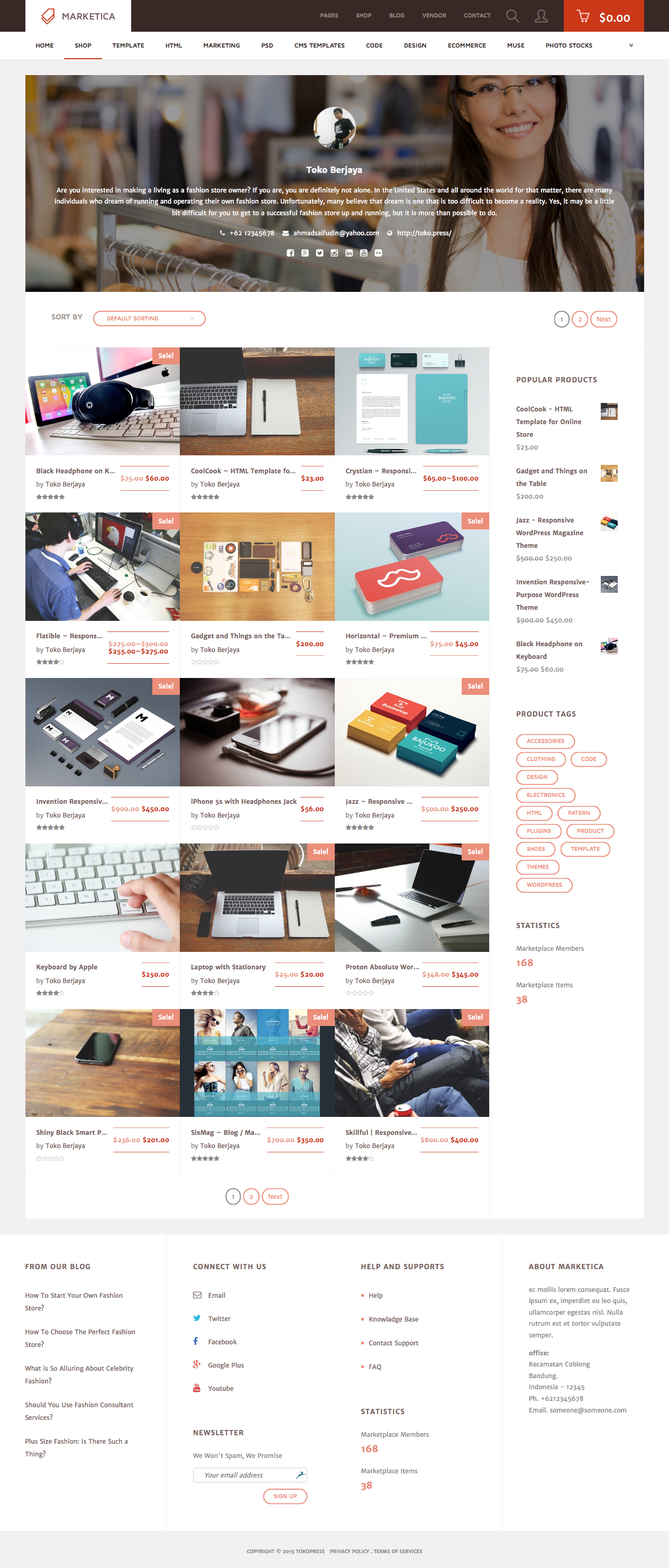
MARKETICA_PREVIEW/22_marketica2_wcvendors_vendor_page.png
MARKETICA_PREVIEW/23_marketica2_wcvendors_vendor_dashboard.png
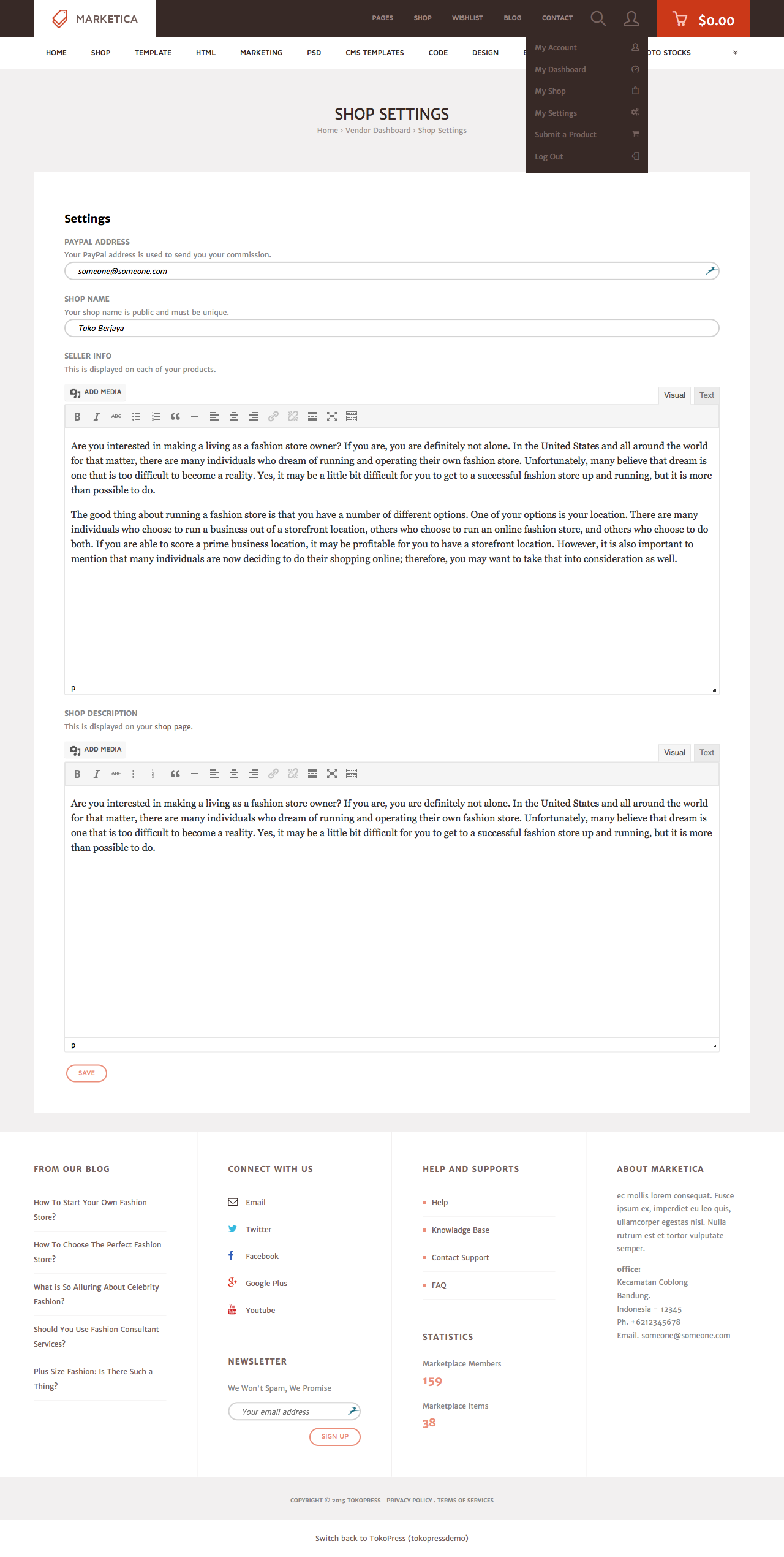
MARKETICA_PREVIEW/24_marketica2_wcvendors_shop_settings.png
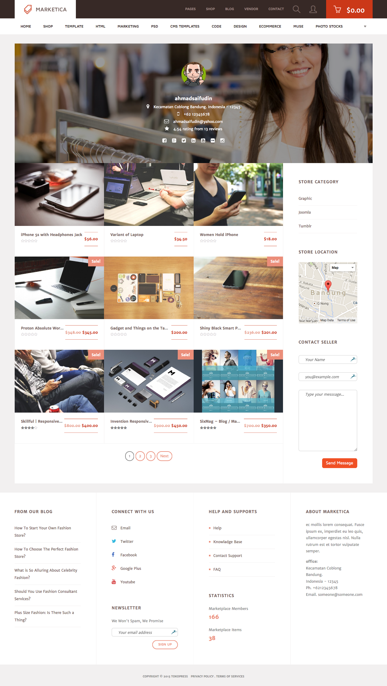
MARKETICA_PREVIEW/25_marketica2_dokan_vendor_store_page.png
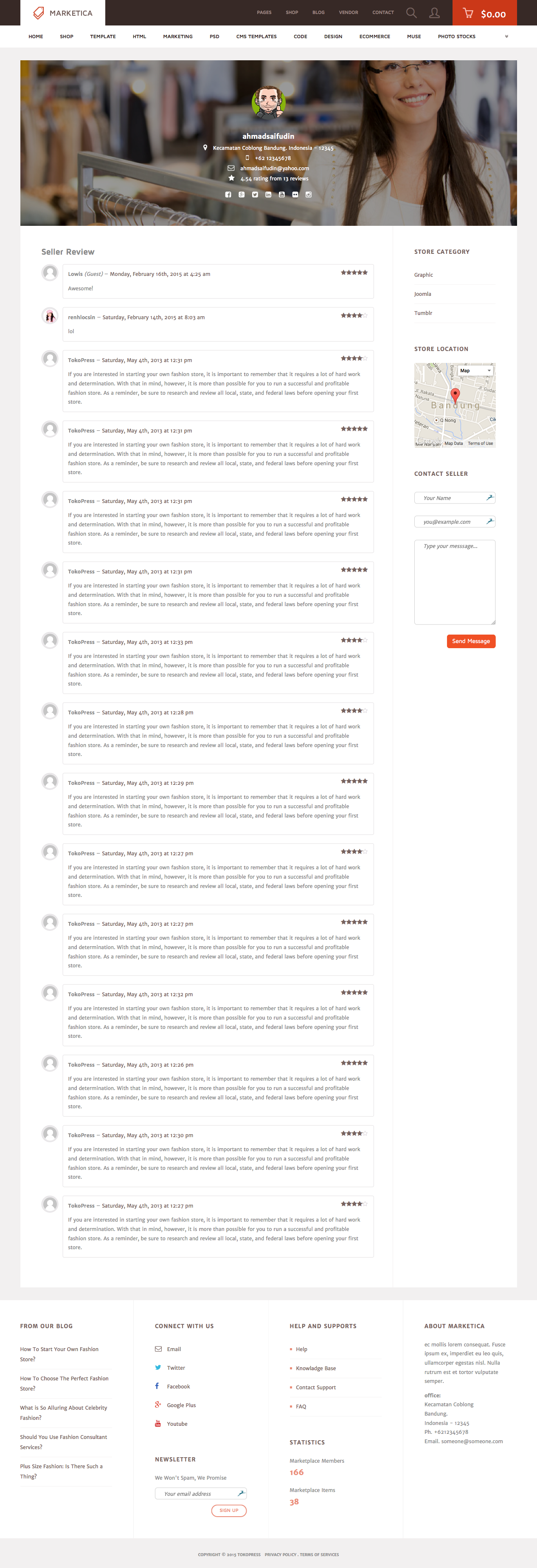
MARKETICA_PREVIEW/26_marketica2_dokan_vendor_review_page.png
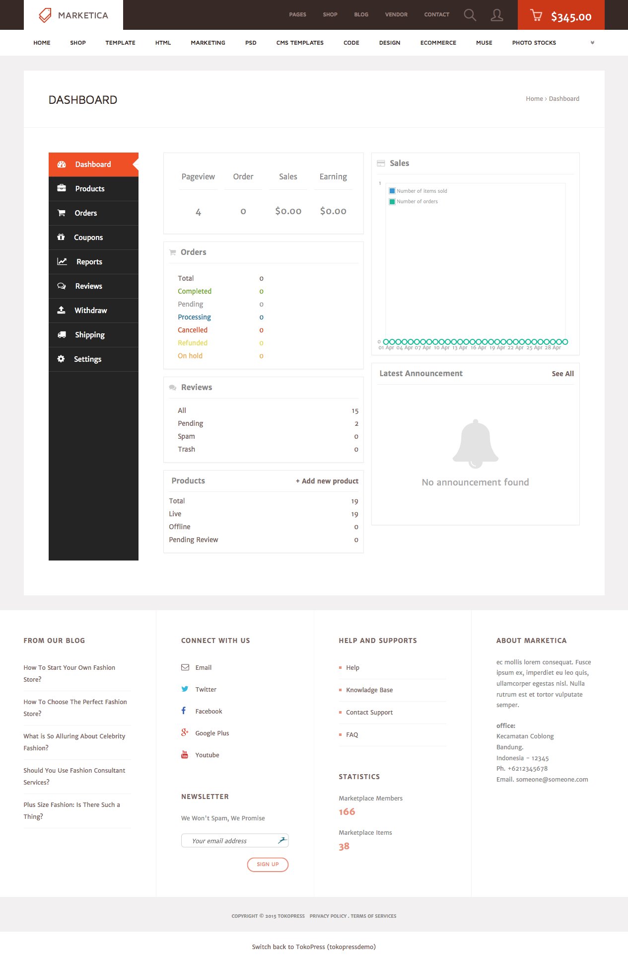
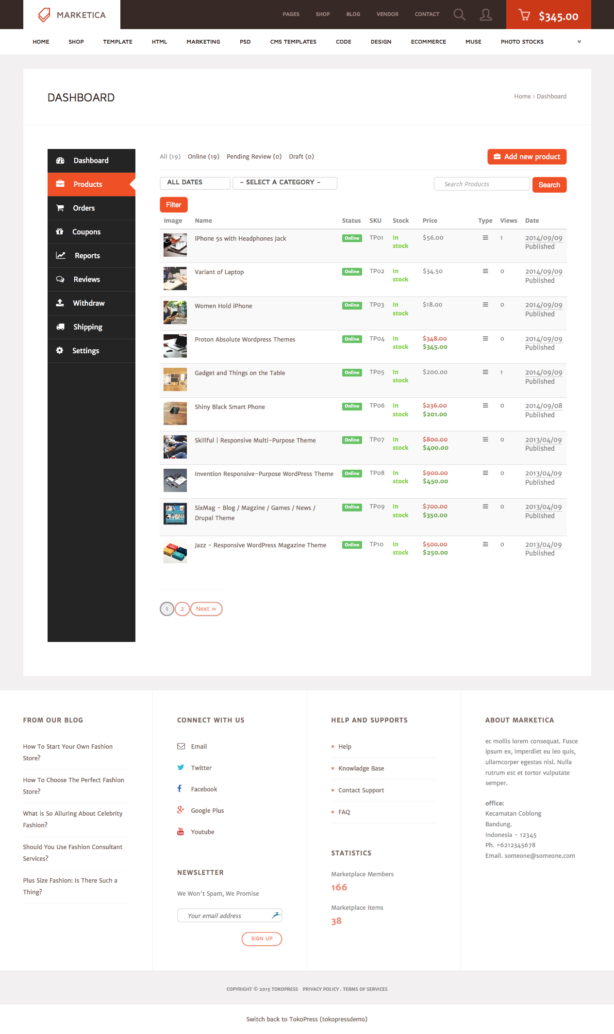
MARKETICA_PREVIEW/27_marketica2_dokan_vendor_dashboard_page.png
MARKETICA_PREVIEW/28_marketica2_dokan_vendor_dashboard_products_page.png
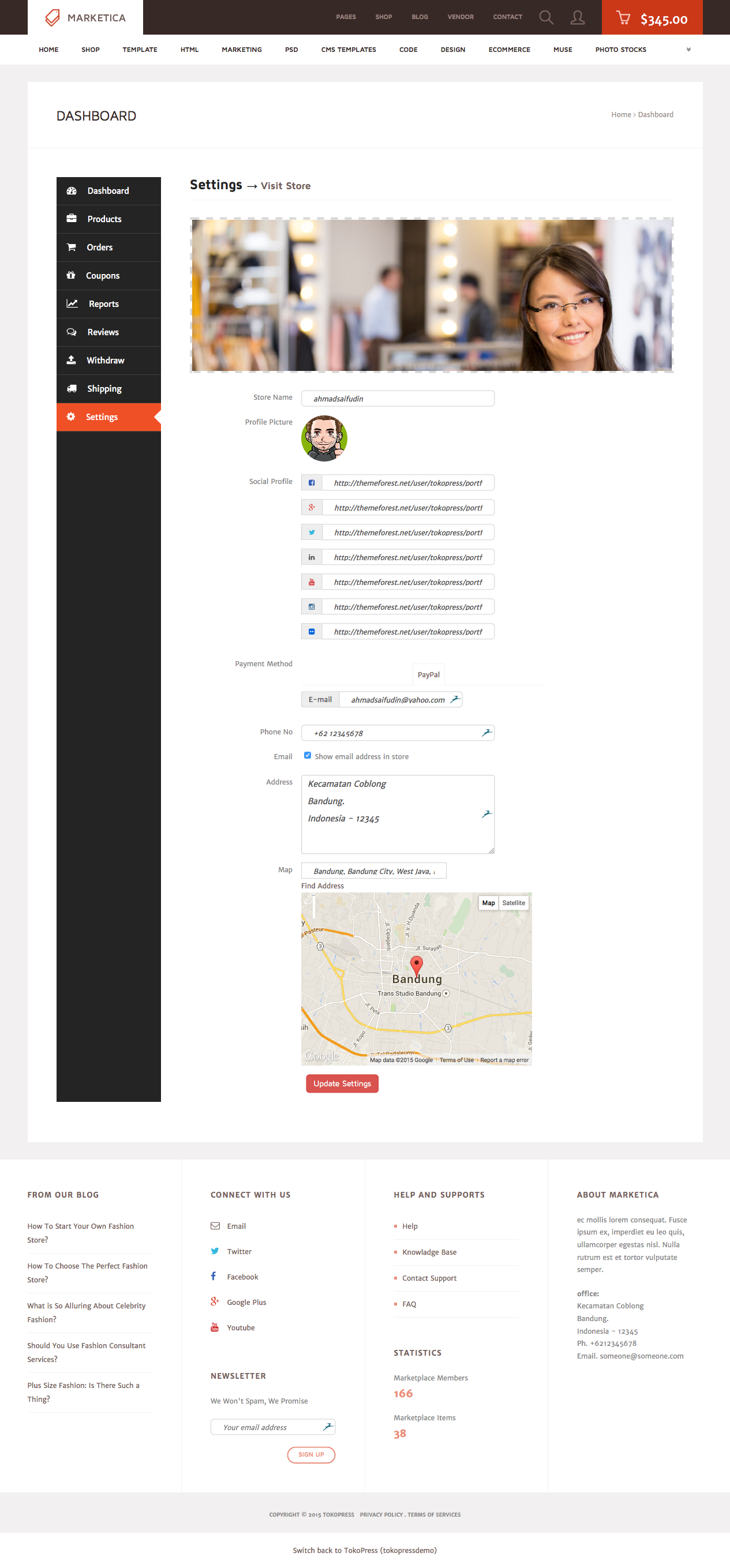
MARKETICA_PREVIEW/29_marketica2_dokan_vendor_dashboard_settings_page.png
FAQ
SLOT88 emang paling ngetop bonus new member melimpah setiap hari, dijamin makin seru dan makin gede peluang menangnya.
Cukup modal receh, asal hoki nempel bisa auto menang terus tiap hari.
Teknologi kekinian banget, dan tim CS-nya selalu on buat bantu lo kapan aja.
Banjir promo dan event tiap hari, bikin lo betah main dan dompet makin tebel.